Meet Sanjakumar Jariwala is currently a postdoc at Biotech Research & Innovation Centre at University of Copenhagen. Former PhD student at DANDRITE, Department of Biomedicine - Neurobiology at Aarhus University.

In my project, I am studying the mechanisms of natural forgetting in the context of learning and memory, with an emphasis on relating these processes to long term potentiation (LTP) and depression (LTD) of synaptic strength. It is well established that the strengthening of synaptic connections is related to memory formation, however very little is known about forgetting.
I was inspired by the concept of the reaching hands from “The creation of Adam” by Michelangelo, and by the canonical representation of a synapse.
More of Andrea Moreno’s artwork is available on: www.amscienceart.com
Andrea Moreno is currently an Assistant Professor at Department of Molecular Biology and Genetics - Neurobiology and affiliated to DANDRITE at Aarhus University.

Mateusz Dyla is currently a Senior Scientist at Novonesis. Former postdoc at DANDRITE, Department of Molecular Biology and Genetics - Neurobiology at Aarhus University.

Mateusz Dyla is currently a Senior Scientist at Novonesis. Former postdoc at DANDRITE, Department of Molecular Biology and Genetics - Neurobiology at Aarhus University.
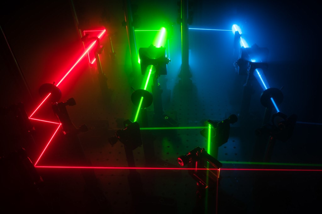
This picture presents a custom-built microscope, equipped with three powerful lasers and accessories controlling their properties, including mirrors to direct laser beam paths. Such microscopes can be used to study proteins labeled with fluorescent dyes. The laser beam paths are typically not visible in the air, so I poured some ‘smoke’ from dry ice sublimation onto the microscope. This resulted in the laser beams being scattered by the small particles suspended in the air, revealing laser beam paths in their full glory.
Mateusz Dyla is currently a Senior Scientist at Novonesis. Former postdoc at DANDRITE, Department of Molecular Biology and Genetics - Neurobiology at Aarhus University.
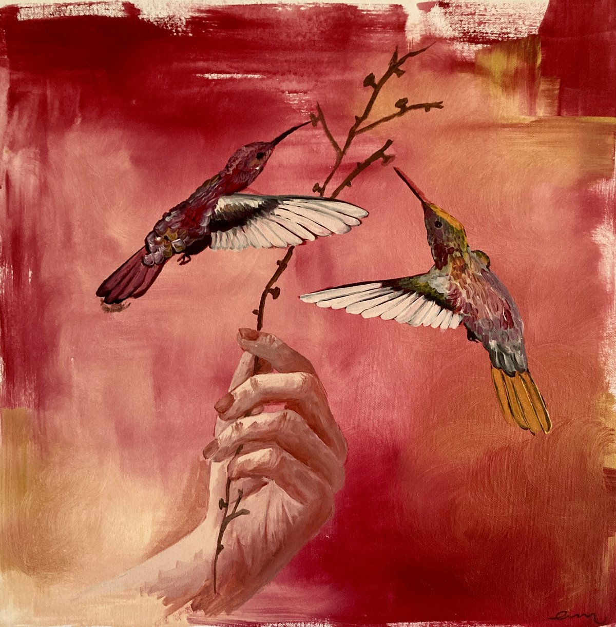
This painting shows a hand holding a dendrite — a branch‑like extension from a neuron. A neuron consists of a cell body, a long axon, and many dendrites, where synapses form and signals are exchanged. In everyday terms, this is where our memories take physical shape: not in one specific place, but as changes in synaptic strength across large networks of neurons.
The hummingbirds represent present‑moment experiences that “feed” the synapses. They symbolize how new information engages with and reshapes older traces. Memory is therefore dynamic: something continually reconstructed, not something fixed. The hand emphasizes both agency and fragility. Our most intimate experiences depend on microscopic structures that can be stabilized, altered, or lost.
Understanding how memories are updated at the synaptic level is important for developing treatments for PTSD, addiction, and neurodegenerative diseases. Made by Andrea Moreno, Associate Professor at Department of Biomedicine, DANDRITE, Aarhus University.

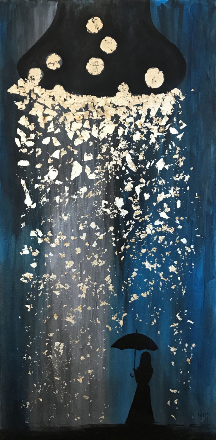
When an idea germinates - an artistic interpretation. The illustration represents the very first beginning of an idea originating from the depth of our brain. Perhaps the idea will perish and be forgotten. Perhaps it will grow bigger, branch out and give rise to new ideas. Perhaps it will grow old and turn into wisdom.
Mette Richner is currently an AC-TAP at the Department of Biomedicine at Aarhus University and holds a PhD in neuroscience. Former Assistant Professor at DANDRITE, Department of Biomedicine - Neurobiology at Aarhus University.
The image represents a presynaptic terminal releasing neurotransmitter contained in vesicles, the effect of which is prevented by blocking arrival to the postsynaptic site. Spontaneous neurotransmitter release could be a way of weakening the synaptic connections that sustain memory.
More of Andrea Moreno’s artwork is available on: www.amscienceart.com
Andrea Moreno is currently an Assistant Professor at Department of Molecular Biology and Genetics - Neurobiology and affiliated to DANDRITE at Aarhus University.
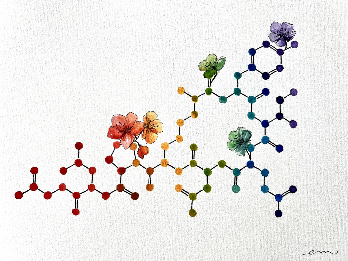
This piece portrays the oxytocin molecule, a small peptide produced in the brain that influences how we form social bonds. In simple terms, oxytocin helps the brain assign emotional significance to social experiences such as motherhood, friendship, romantic attachment, and trust.
The molecule is painted in rainbow colours to emphasize the diversity of attachment and love. The surrounding flowers symbolize synaptic plasticity – the brain’s capacity to change. Emotional memory, like a blooming flower, emerges only under precise biological conditions. Understanding how different molecules influence memory has implications for treating PTSD, addiction, and neurodegenerative diseases. It highlights that memory is biological – and therefore modifiable.
Made by Andrea Moreno, Associate Professor at Department of Biomedicine, DANDRITE, Aarhus University.
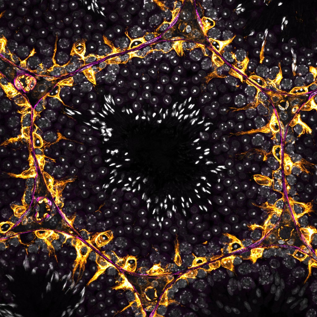
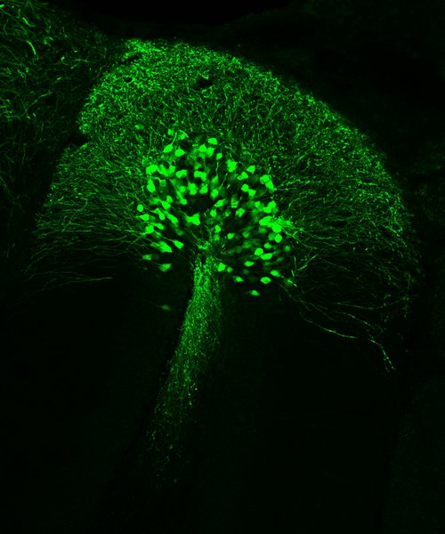
Sperm is created in the seminiferous tubules of the testis via the sequential process of spermatogenesis, which converts germ cells into mature sperm containing concentrated nucleic matter, which holds genomic information. This process is dependent on the work of Sertoli cells within the tubules, which support and nourish the developing sperm cells. In this image, individual tubules are surrounded by basal lamina layers stained for smooth muscle actin (purple), Sertoli cells are stained for vimentin (yellow), and sperm cells and progenitors are identified by nuclear marker DAPI (white). From the edge of each tubule into its centre, it is possible to see the stages of spermatogenesis progress, culminating in an inner layer of bright white sperm cells ready to be shed: a circle of pre-life. The section was stained via immunohistochemistry and imaged on a Leica STELLARIS 8.
Silke Blair Chalmers is currently a postdoc at Department of Biomedicine – Neurobiology at Aarhus University.

The painting portraits a synaptic connection between brain cells that is about to “bloom” to transmit a neural impulse. You can observe a synaptic bouton, which consists of presynaptic and postsynaptic terminals. These terminals contain various proteins, sorting vesicles, and organelles that are continuously transported between the cell soma and the synaptic terminals. The fish symbolizes actin filaments and their structural dynamics, which is an active cytoskeletal system critical for synapse remodeling, plasticity, and signal transmission. Synapses are fundamental communication sites between brain cells. They are indispensable for all aspects of the brain function including learning and memory. Impaired synaptic biology can have detrimental consequences, leading to various neurological and psychiatric disorders, or even brain death.
Alena Salasova is currently an Assistant Professor at Department of Biomedicine – Neurobiology at Aarhus University

This light-sheet microscopy image captures an intact mouse embryo frozen in time and space. The fluorescent signal highlights every single motor neuron in the organism. Motor neurons are specialized cells that transmit impulses between the brain and muscles, allowing us to move, speak, and smile. By making the embryo transparent, we can trace and reconstruct motor nerve growth in 3D with high precision. Indeed, this approach enabled us to visualize developing motor neurons in intact wild-type and mutant embryos, allowing us to image, reconstruct and measure 19 distinct motor nerves with a precision of a few micrometers. Our findings were recently published in the scientific journal Cell Reports (https://doi.org/10.1016/j.celrep.2023.113333).
Embryonic development requires a detailed roadmap that guides cells to their final destinations, roles, and functions. Understanding these processes can help develop new therapies for various neurological disorders and spinal cord injuries.
Josef Lavicky is currently working at Masaryk University, Faculty of Medicine, Department of Histology and Embryology; Brno Czech Republic. Alena Salasova is currently an Assistant Professor at Department of Biomedicine – Neurobiology at Aarhus University

Brain cells are called neurons and function like small computers in the brain, helping us think, feel, and move. By growing nerve cells in a dish, we can study human brain diseases — and deeper insight is essential for developing new treatments. A human cell model allows us to investigate disease processes in an environment that more closely resembles human biology than animal models do.
In the image, two different cell types are labelled with fluorescent proteins so they can be visualized under the microscope. Human cortical neurons (in purple) are grown together with rat astrocytes (in green), which help them thrive. All cell nuclei are stained blue with a DNA dye — the nucleus is the cell’s “control room, ” where the genetic material is stored and many of the cell’s key processes are regulated.
In our laboratory, we study the molecular mechanisms underlying Parkinson’s disease, and my role is to develop a human cell model that can help us understand these processes more clearly.
Made by Anissa Hammi, PhD student at Department of Biomedicine, DANDRITE, Aarhus University.

This microscopy image shows human cells grown in the laboratory. The blue areas mark the cell nuclei, while the purple colour highlights the cell’s internal skeleton. Inside one of the nuclei, a thin green thread‑like structure can be seen. It consists of the protein alpha‑synuclein, a naturally occurring protein produced by all humans. In Parkinson’s disease, alpha‑synuclein can change shape and clump together into fibers and aggregates that disrupt the cell’s normal function.
My research investigates how alpha‑synuclein changes structure, accumulates in cells, and affects their biology. By recreating these protein accumulations in cell models, we can study the early disease processes under controlled conditions. A cell model is a cultured collection of cells that functions as a simplified and controllable version of living tissue. The goal is to understand how and why these protein structures form — and ultimately contribute to the development of treatments that can slow or prevent Parkinson’s disease and related disorders.
Made by Hjalte Gram, who is a Scientist at PACE - Lundbeck Foundation Parkinson's Disease Research Center, and former PhD student at DANDRITE.

This microscope image shows structures of the mature mouse mammary gland: a complex network of branching ducts. These ducts form the developmental “roots” that determine the breast’s future ability to produce milk and nourish the next generation – much like the roots of a tree shape how well it will flourish in spring. Specialised cells, shown in green, sense and respond to hormones, allowing the tissue to change throughout the menstrual cycle. These changes prepare the breast for the dramatic transformation required during pregnancy and breastfeeding.
By understanding the foundational biology of breast tissue prior to pregnancy, we hope to uncover the causes of breastfeeding challenges such as low milk production. Improving breastfeeding outcomes supports not only infant and maternal health, but also the wellbeing of families and communities worldwide.
Made by Laura Bruus Bjerre, PhD student at the Department of Biomedicine, Aarhus University, affiliated to DANDRITE
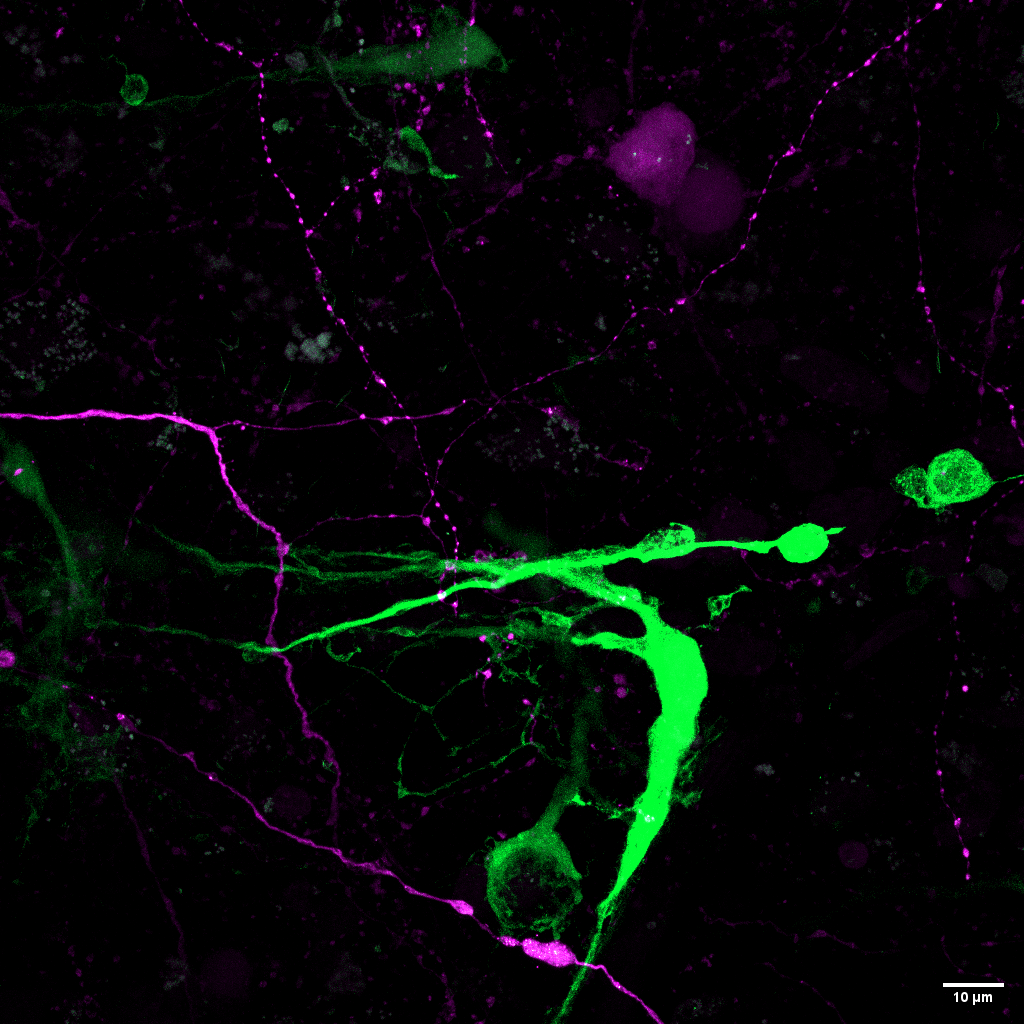
In magenta, a healthy brain cell – a neuron – extends its intricate network of projections across the image. In green, a glioblastoma tumor cell appears, representing one of the most aggressive forms of brain cancer.
Where their structures come into close contact, an important biological interaction takes place. By studying how healthy brain cells and tumor cells communicate, we hope to identify new therapeutic targets that could improve treatment options for patients facing this devastating disease.
Made by Markus Ørnsvig Christensen, Master’s student in Molecular Biology and part of DANDRITE, Aarhus University
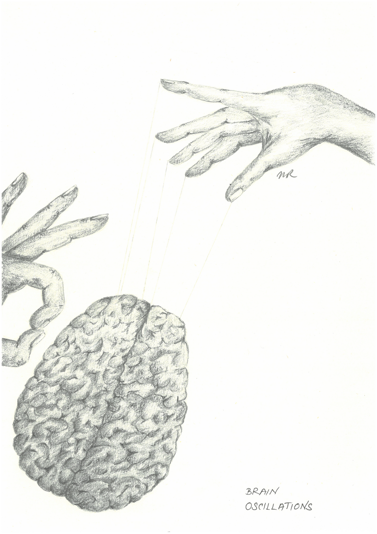
What sets the very first thought in motion? The moment the brain wakes up, neurons begin firing in sync, and a small electrical wave shoots through the network. When many of these waves arise at the same time, they become brain oscillations — the brain’s own rhythms that help shape every thought, decision, and sensation.
But what makes the brain oscillate? Neurons send tiny electrical nudges to one another, like rhythmic signals in a shared circuit. And then the world intervenes: a sound, a flash of light, a touch, a gust of cold air. Each new input gives the rhythm a push and makes the brain adjust the tempo, strength, or pattern of its oscillations so we can respond quickly and meaningfully.
Oscillations are the brain’s way of staying organized — a constant dance of electrical rhythms that makes thought possible and never stops.
Made by Mette Richner who is Senior Researcher at HemeLab, Department of Hematology at Aarhus University Hospital. Formerly employed as an assistant professor at DANDRITE.

Ever wondered what’s going on inside your brain? Picture a forest. A wild, living landscape with billions of trees waving their branches, all trying to talk at once. Congratulations – you’re now inside the Dendritic Forest. The trees stretch out their branches and whisper messages to one another. Sometimes they even shout: “HELLO? ANYONE? I HAVE A SIGNAL!” Tiny electrical sparks shoot through the woods – nature’s own version of texting.
Some parts are beautiful: diverse trees working together, sharing signals, growing ideas, storing memories. Some are dense and confusing: tangled thickets where thoughts get lost, like when you walk into a room and forget why. And the dark corners? That’s where your brain stores all your “I should really remember this” plans. But there is always light. Always movement. Always connection. Every time a neuron fires, a spark cuts through the shadows and guides the way. Welcome to the Dendritic Forest – where your brain works hard, looks confusing, and occasionally forgets your keys.
Made by Mette Richner who is Senior Researcher at HemeLab, Department of Hematology at Aarhus University Hospital. Formerly employed as an assistant professor at DANDRITE
Neurotransmission is the brain’s way of passing messages from one neuron to the next – like a relay race, but with far more drama and much better choreography. When a brain cell – a neuron - wants to speak up, it releases neurotransmitters: tiny chemical messengers that leap across the synapse, the gap between two neurons, each with its own personality. Some rush across at full speed shouting, “Move, I’ve got urgent information!” Others float gracefully like ballet dancers delivering a whispered secret.
And then there are the tip‑toe types, quietly sneaking their message across without disturbing anyone. Together, this eclectic cast keeps your thoughts flowing, your mood steady, your movements coordinated, and your inner monologue… well, occasionally questioning your life choices. Neurotransmission is a microscopic dance party that never stops, because neurons always have something to say.
Made by Mette Richner who is Senior Researcher at HemeLab, Department of Hematology at Aarhus University Hospital. Formerly employed as an assistant professor at DANDRITE.
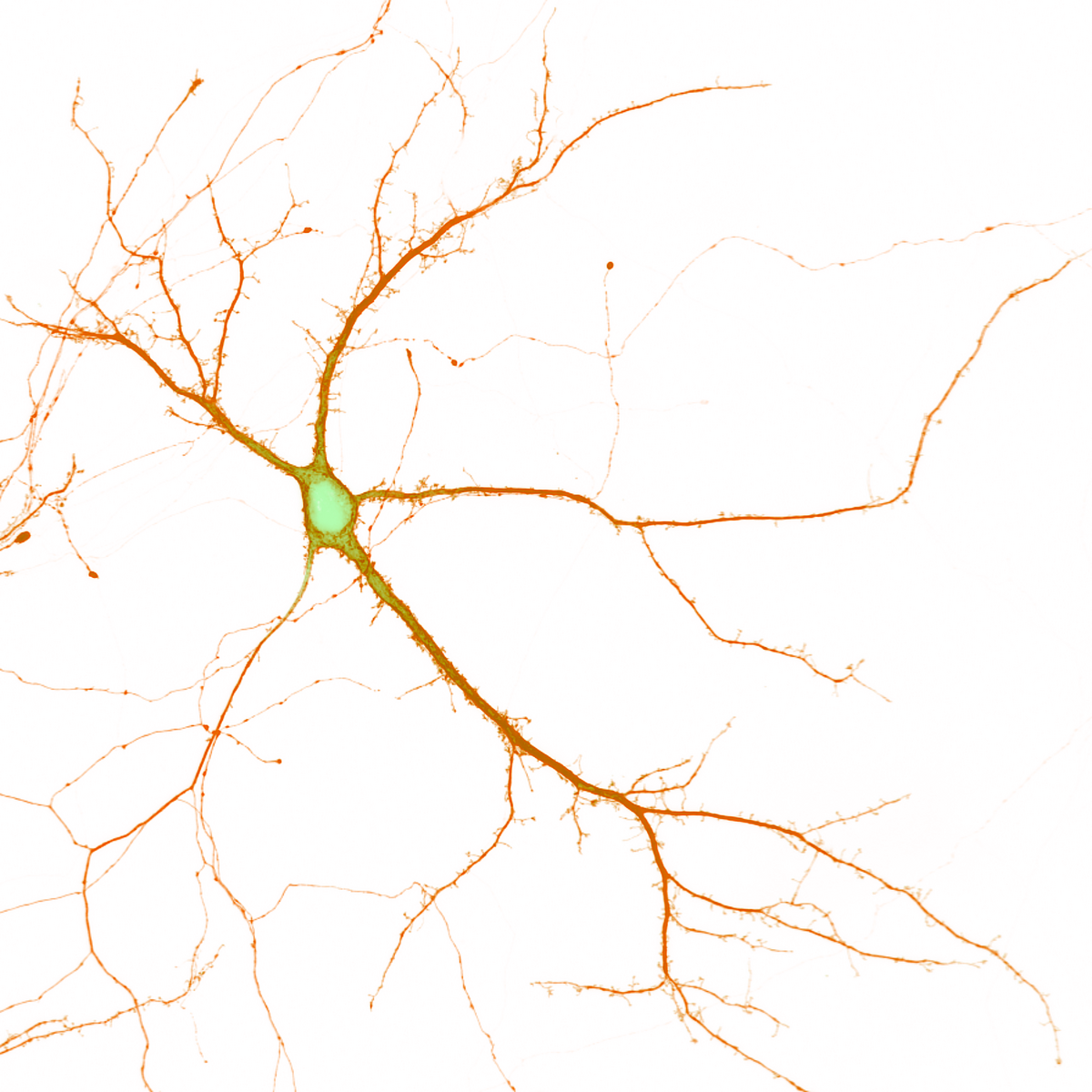
The image shows an isolated neuron — a brain cell consisting of a cell body, long branching extensions, and a network of connections that allow it to send and receive signals. In this cell, internal stress is marked in green, and my goal is to understand how neurons cope with this kind of strain.
In this experiment, I intended to visualize stress in different parts of the neuron. But due to a technical error, I was only able to highlight stress on the cell’s surface, shown in orange. This unexpected limitation made me think of a human parallel: how periods of isolation — suggested by the white background — can amplify the stress we carry inside.
Neurons are built to reach out and communicate with one another. Perhaps we can learn from them and reach out when we ourselves feel under pressure.
Made by Vivek Sanjay Belapurkar, Postdoc at DANDRITE, Department of Molecular Biology and Genetics, Aarhus University
