New viral tool for mapping and imaging brain activity
Researchers from Nabavi and Yonehara Labs have developed an improved method that can map the connections between neurons.


Understanding how the brain works and how different parts of the brain communicate is essential for comprehending the development and occurrence of diseases and degeneration in the brain.
Now, researchers from Nabavi and Yonehara Labs have developed an improved method that can map the connections between neurons.
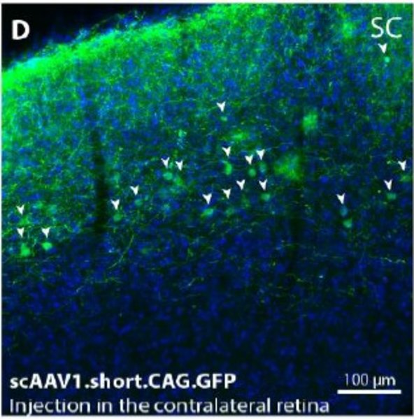
By employing a viral vector known as AAV1 to anterogradely label synaptically connected brain cells, the researchers have determined that this vector is significantly more versatile. This is because it can be used in the brain of any mammalian animal model in an unbiased and simpler manner.
Islam Faress, postdoc and the lead author highlights that “this new method not only aids in our understanding of how brain cells are interconnected but can also be employed for mapping and imaging brain activity”.
The method is accepted to be published in Molecular Brain as a brief communication under the title: “Recombinase-Independent AAV for Anterograde Transsynaptic Tracing”.